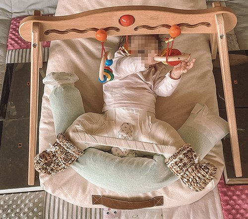
Open (surgical) reduction of both hip joints in an infant
Treating congenital hip dislocation with surgery has become quite the rarity. This is due to the fact that we perform ultrasound screening using the Graf method after birth as standard in most cases and because of the possible conservative early treatment options at our disposal. In some cases, however, the femoral head cannot be positioned into the socket by non-surgical means, meaning surgical treatment becomes necessary.
Our case study shows the development in a female infant. After preliminary treatment at another paediatric orthopaedic centre was unfortunately unsuccessful, the parents brought their child to our Paediatric Orthopaedics department. An attempt had been made previously to stabilise the hip joints using traction, two closed reductions under anaesthesia and the application of a plaster cast. Given the previous unsuccessful courses of treatment, the parents were particularly worried.
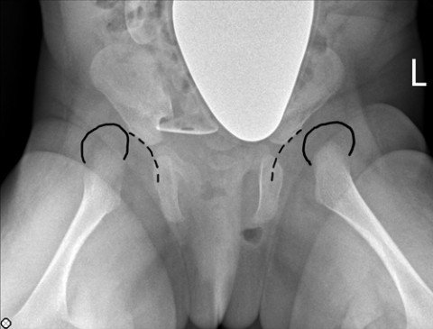
When the infant presented to us at the age of two months, both the X-ray and the ultrasound continued to showed significant dislocation of the femoral heads (Figure 1). We attempted another treatment method involving a Pavlik harness. We did, however, come to the realisation that, although the femoral heads could be brought into a better position in front of the opening to the acetabulum, there was an obstruction, which prevented the femoral head from assuming the correct position inside the acetabulum.
The obstruction preventing reduction required surgery.
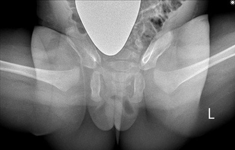
At the age of four months, we first performed an arthrogram (examination using contrast medium) of the hip joints in a well-prepared operation accompanied by our highly specialised paediatric anaesthesia team. This confirmed the suspicion of a soft tissue interposition preventing reduction. We therefore immediately performed a careful open reduction of the hip joint under the same anaesthesia. The soft tissue interposition was lifted out of the joint and the joint capsule was closed so as to provide very good stability. The X-rays and the MRI performed without anaesthesia while the patient was in the postoperative plaster cast showed excellent positioning of both femoral heads in the acetabulum (Figure 2).
Further examinations also showed an optimal result and the girl recovered very quickly from the operation (Figure 3).