Featured Cartilage chip implantation (minced cartilage): the neat and gentle cartilage repair technique
A newly developed cartilage repair procedure has shown promising results in terms of repairing damage to knee cartilage. Cartilage chip implantation involves mechanically crushing the patient’s own cartilage tissue into “chips”. These are mixed with the patient’s own blood, which has been prepared beforehand, and then reused. The advantage of this approach compared to other cartilage repair procedures is that only one operation is required, which is less strenuous for the patient and takes less time. With its use of endogenous substances, the procedure offers promising potential.
Injuries and damage to articular cartilage are becoming increasingly common. Cartilage damage usually has a restrictive impact on a patient’s sporting activities in particular. Osteoarthritis may develop gradually.
Previously, cartilage was usually repaired over the course of two operations
One treatment option is autologous chondrocyte transplantation. With this method, an initial operation is carried out to remove the body’s own (autologous) cartilage cells (chondrocytes). These then have to be propagated over several weeks in a special cell culture laboratory, after which a second operation is performed to insert them back into the knee joint. This procedure has its disadvantages, however, such as changes to the cells occurring during the laboratory phase, the resource-intensive cultivation process with stringent regulatory requirements and the high costs arising from this.
Arthroscopic minced cartilage chip implantation (also known as autologous cartilage chip transplantation or autologous cartilage repair) offers a simpler and more cost-effective alternative.
The minced cartilage implantation (MCI) method (cartilage chip implantation)
At Schulthess Klinik, we have been using the minced cartilage implantation (MCI) procedure for over five years now, alongside other cartilage repair methods. Our aim in doing this is, on the one hand, to try to reduce the pain caused by the cartilage damage and, on the other hand, to prevent the development of osteoarthritis of the knee.
Since the surgeons require fibrin at a later stage, they take a blood sample at the beginning of the operation. Fibrin is a protein produced during blood clotting due to the effects of thrombin (the enzyme that causes blood to clot) and PRP (platelet-rich plasma). It acts as an endogenous adhesive and, in the case of the MCI procedure, it helps to fix the cartilage chips in place. PRP, meanwhile, is very effective at promoting the propagation and development of cartilage cells. It also has positive effects on coagulation (less bleeding during and after surgery) and inflammation (irritation resulting from cartilage damage and surgery) in the joint as a whole.
An all-in-one operation
The surgeons can perform the technique openly or arthroscopically (via a small incision using an endoscope/camera). Most procedures are now carried out arthroscopically. First, the surgeon removes the damaged cartilage tissue using a small sharp spoon or a ring-shaped curette. They then collect some healthy cartilage tissue from the edge of the damaged area. This cartilage is then crushed immediately after extraction using a special machine and mixed with PRP obtained at the same time. This produces a “paste”, which can then be inserted easily and neatly into the prepared area where the cartilage was damaged. Finally, the thrombin produced from the PRP shortly beforehand (also using another special device) is drizzled over the paste. This immediately binds with the PRP, which then produces fibrin (adhesive). The fibrin hardens instantly, thus fixing the transplanted chips in place in the damaged area.
Within a short time (hours), the cartilage cells then begin to divide, anchor to the surface of the damaged area and the surrounding cartilage and build up tissue between the cells. The fibrin automatically breaks down again within a few weeks, allowing the cartilage to build up without any hindrance.
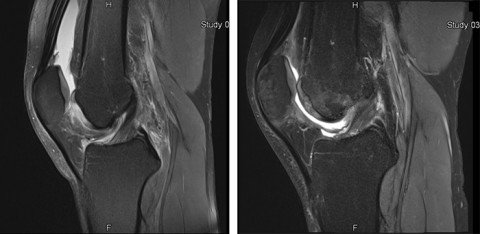
Right: Postoperative MRI image of the same cartilage damage six months after cartilage chip surgery using an arthroscopic technique, showing excellent restoration of the cartilage layer and very good integration of the graft into the surrounding area
Advantages of cartilage chip implantation
Implanting crushed cartilage offers various advantages. For one thing, it only requires one operation and therefore saves both time and costs compared with other procedures. In most cases, the procedure involves a minimally invasive arthroscopic technique and can also easily be combined with any other procedures required at the same time. Cartilage chip implantation is therefore very kind on the patient.
Moreover, this approach also offers promising biological potential, as substances produced by the patient’s own body are removed, processed and immediately reused. Adding the PRP also brings a significant natural catalyst into play – a feature that is currently only found in very few cartilage repair techniques. While the effectiveness of cartilage chips has been acknowledged for decades and has been described in numerous experimental and animal studies, the latest studies show highly promising clinical results.
The MCI procedure is suitable for both minor and extensive cartilage damage as well as cartilage bone damage.
About the arthroscopic minced cartilage implantation (MCI) study
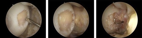
Centre: The same cartilage damage after preparation is complete and the joint has been dried out in preparation for applying the cartilage chips
Right: After the cartilage chips have been inserted arthroscopically and fixed in place with thrombin/fibrin, the final examination with a hook probe reveals a stable cartilage graft.